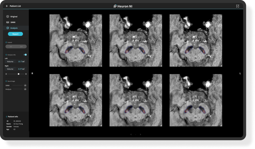
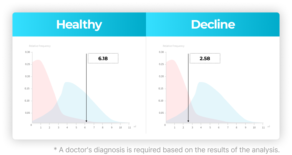
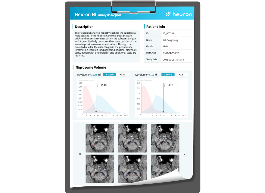
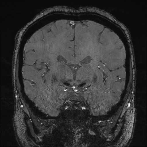
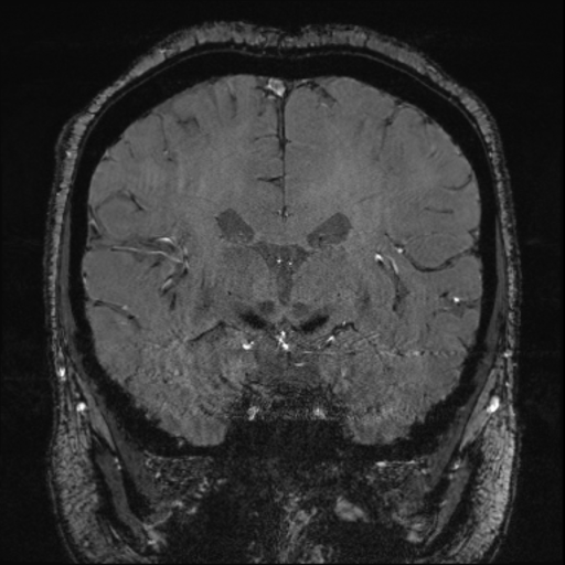
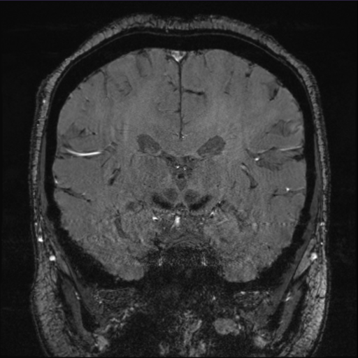
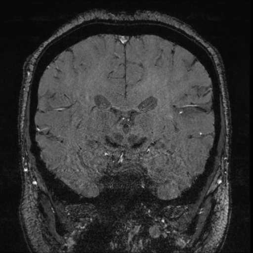
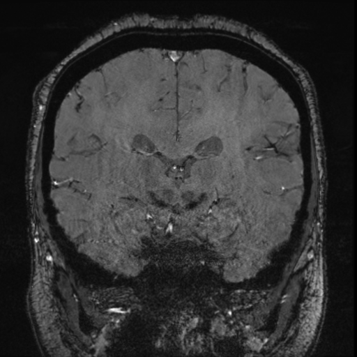
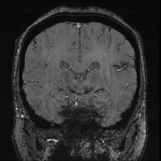
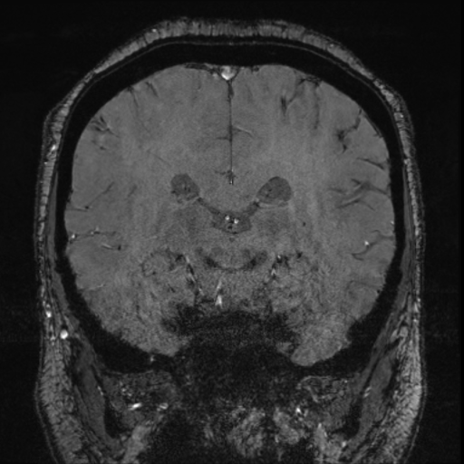
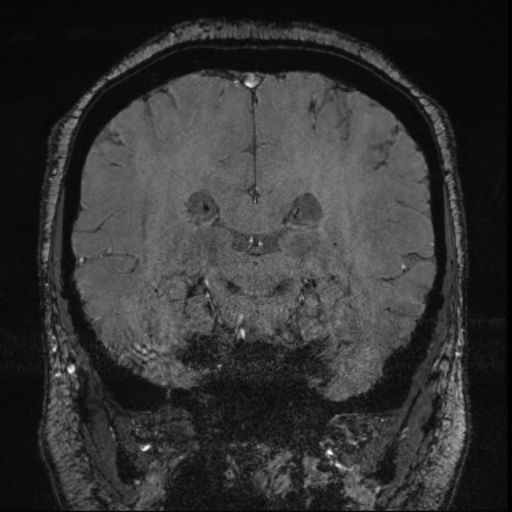
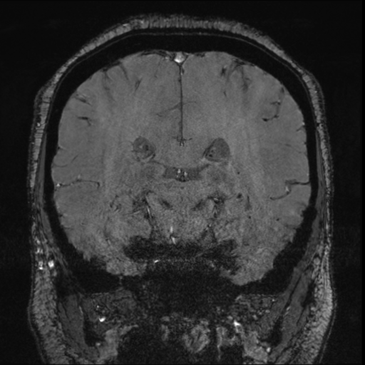
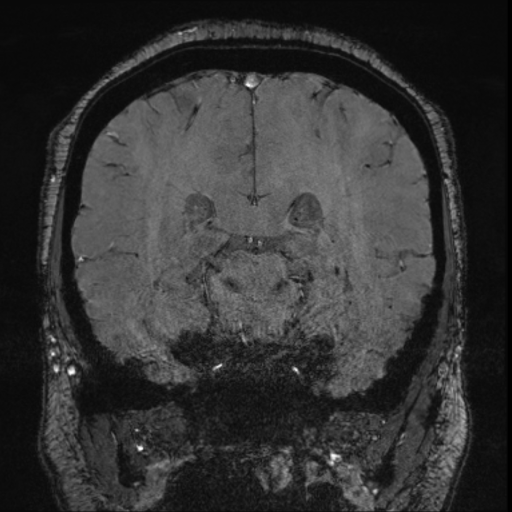
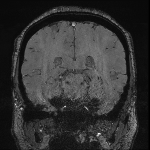
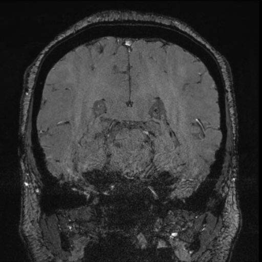
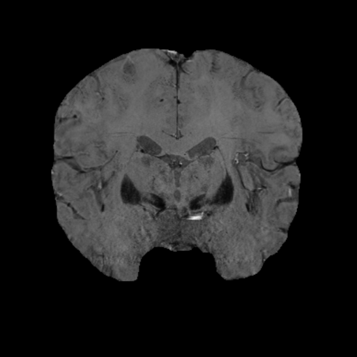
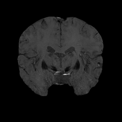
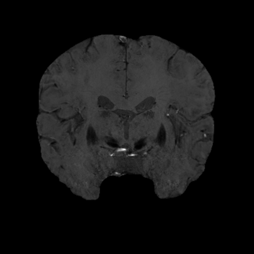
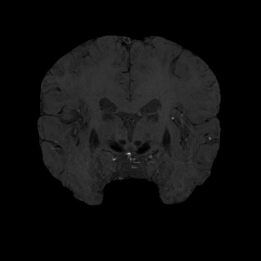
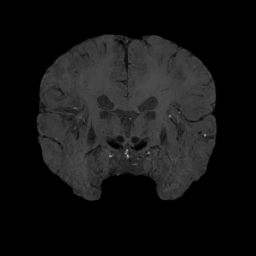
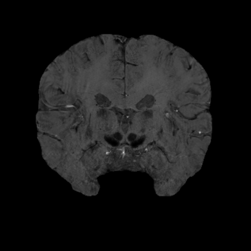
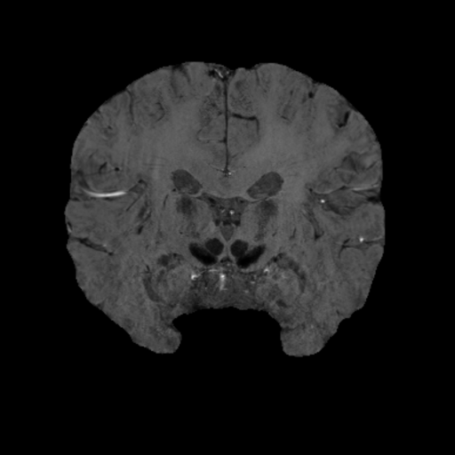
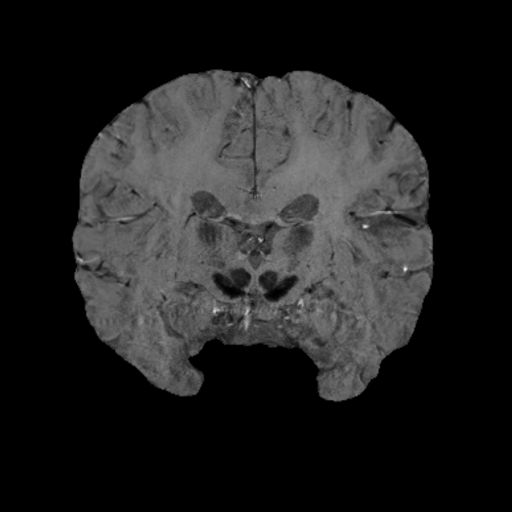
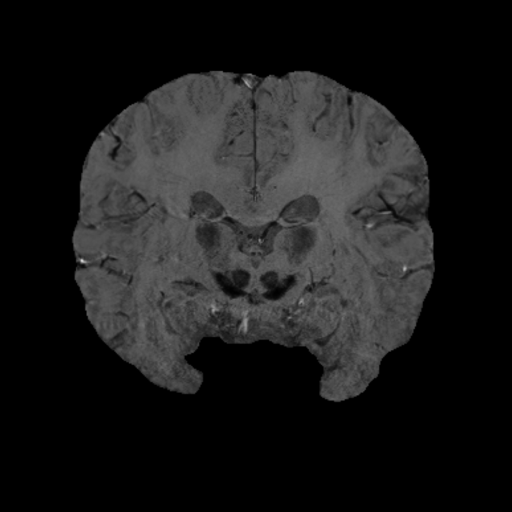
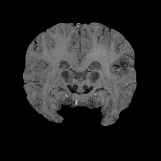
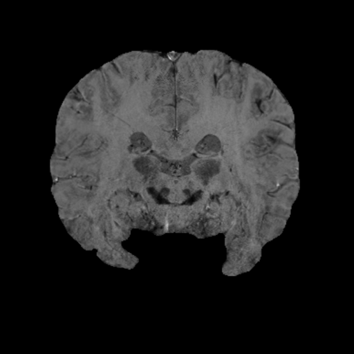
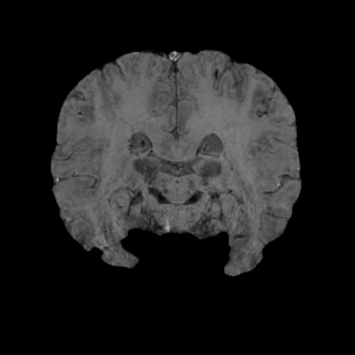
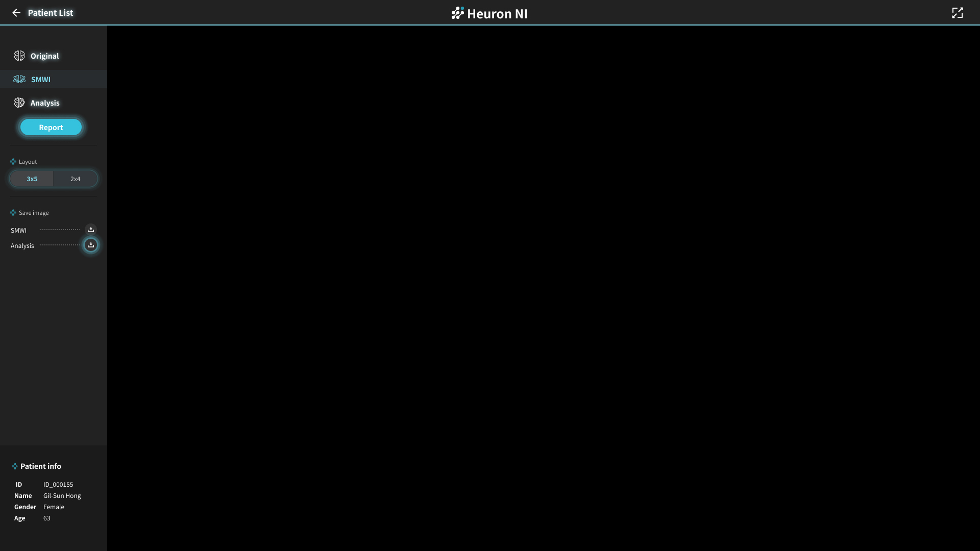
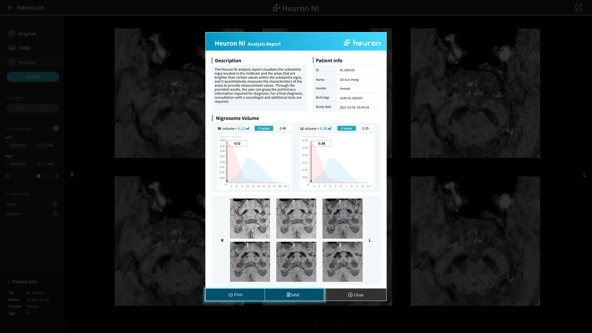
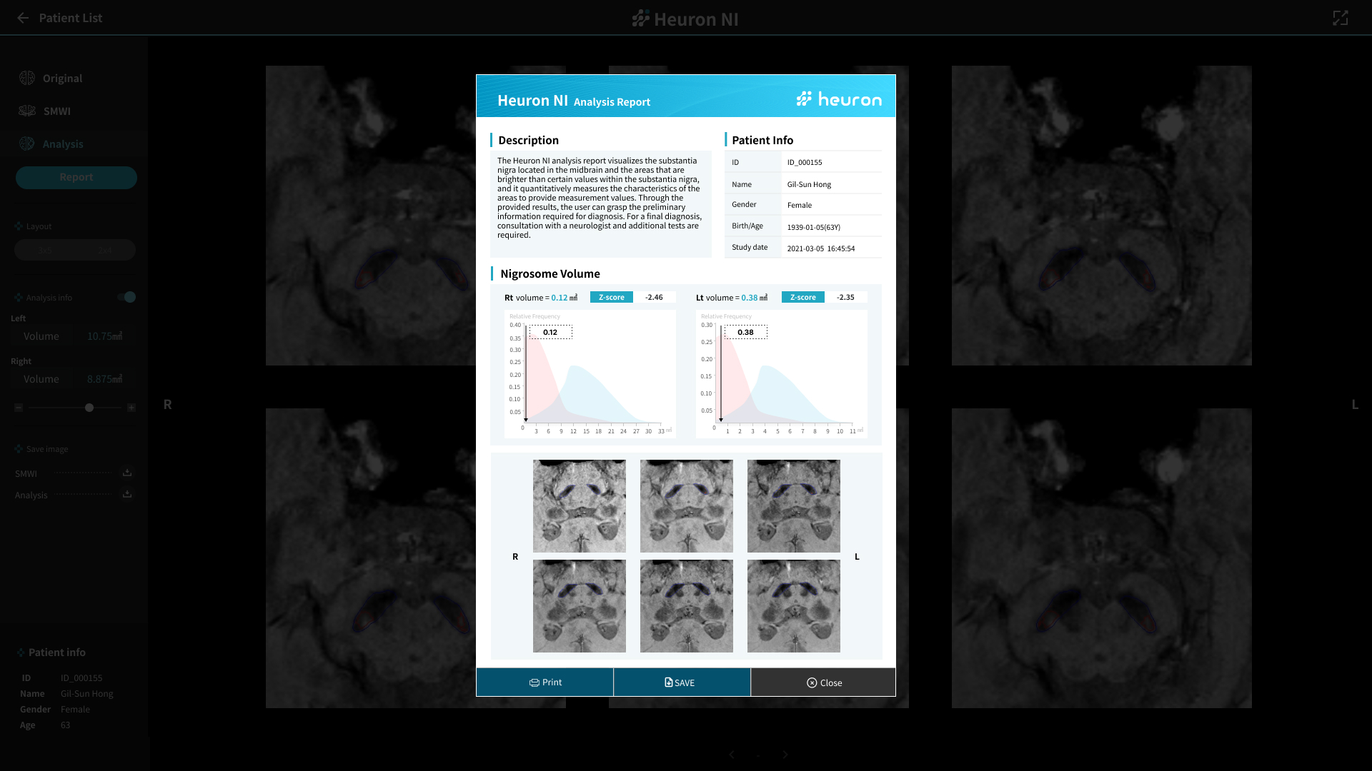
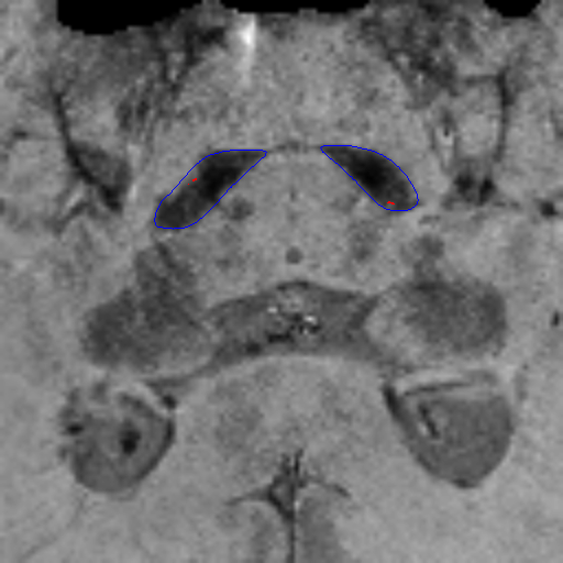
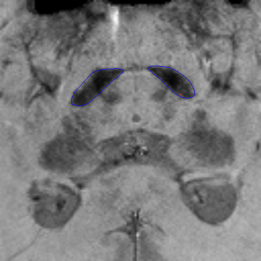
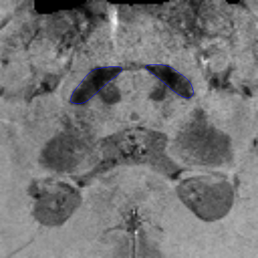
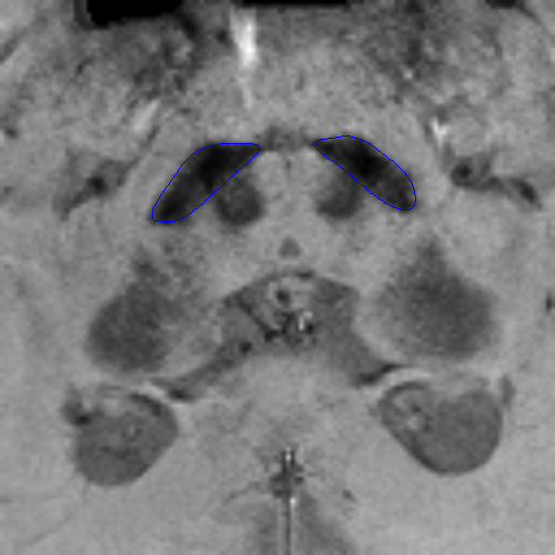
Scroll Down

  • Home

  • >
  • Neurodegenerative

  • >
  • Heuron AgingCare Suite™

  • >
  • Heuron NI

Heuron NI

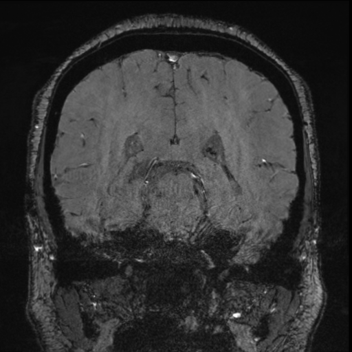
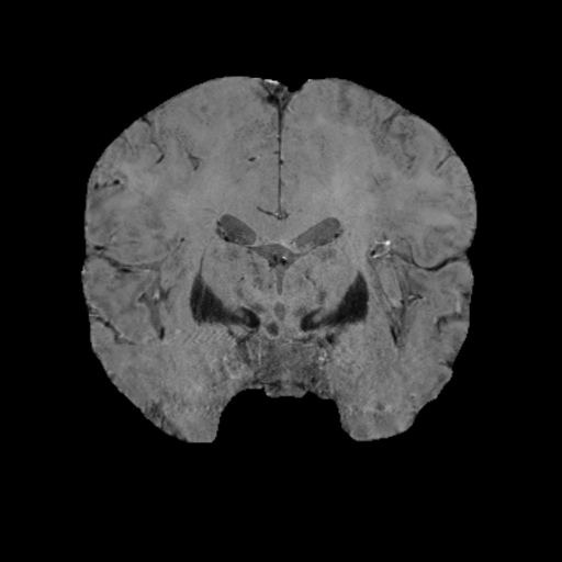
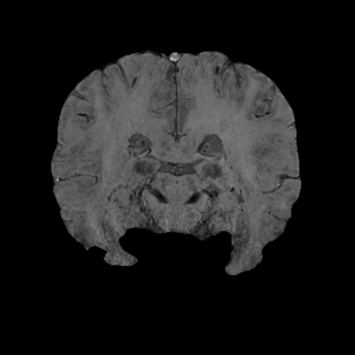
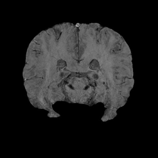
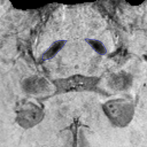
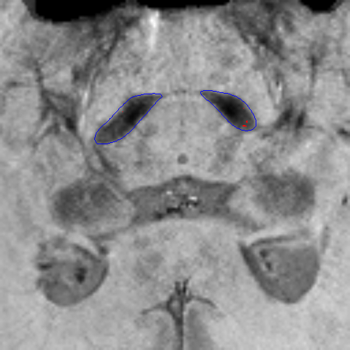
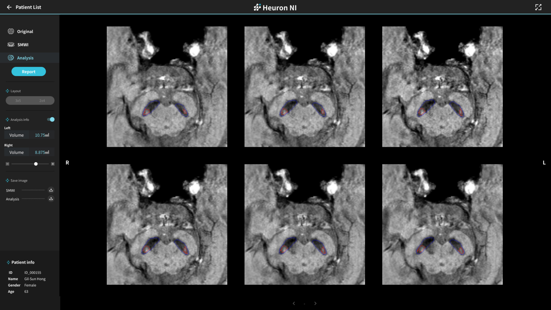
Visualizes and quantifies
Parkinson’s disease lesions.
MFDS Approved

Heuron NI Brochure

  • MRI Analysis

  • Assessment in the
    asymptomatic stage

  • Easy and convenient
    analysis report

Workflow Improvement

Visualizes the nigrosome area on brain MRI and quantitatively measures its volume, assisting in Parkinson's disease diagnosis.

Differentiation of Parkinson’s from other movement disorders that do not exhibit the loss of dopaminergic neurons.

Supported Features

Automatically outputs Z-scores and graphs to indicate the presence of the nigrosome area volume reduction.

Decreased nigrosome area volume indicates a higher possibility of dopaminergic neuron damage, requiring continuous monitoring and observation.

Heuron NI Report

Facilitates doctor-patient communication with a visualized analysis report for more effective follow-ups.

Heuron NI

Nigrosome Quantitative Analysis

Experience the product's features firsthand from your screen.

L
R

Want to learn more about Heuron NI?
Explore more with our demo.

TOP